
The study describes morphometric changes in the adenohypophysis during sudden cardiac death. The study investigated the morphometric parameters of pituitary gland tissues obtained from 75 individuals who died of sudden cardiac death, categorized according to the WHO age classification into cohorts of 18–44, 45–59, 60–74, and 75–90 years and older. The specimens were examined at the Andijan branch of the Republican Scientific and Practical Center for Forensic Medical Examination during the period of 2015–2024. In acute cardiac death associated with ischemic heart disease (IHD), age-specific features revealed that in the 18–44 and 45–59 age groups, the diameters of acidophilic cells, thyrotroph cells, corticotroph cells, and gonadotroph cells significantly increased; these indicators explained the enhancement of metabolic processes and morphofunctional activity in them. In the 60–74 and 75–90 age groups, a statistically significant decrease in the diameters of acidophilic cells, thyrotroph cells, corticotroph cells, and gonadotroph cells was observed, which manifested a decline in cellular metabolic processes. A decrease in the area occupied by the adenohypophyseal parenchyma accompanied by an increase in the stromal area occurred in parallel with advancing age, demonstrating a decline in the gland's morphofunctional activity. These changes were explained by the fact that age-related atrophic and sclerotic alterations of the pituitary gland in these individuals were accelerated against the background of IHD, and atrophic changes accelerated the aging processes.
| Published in | American Journal of Clinical and Experimental Medicine (Volume 14, Issue 4) |
| DOI | 10.11648/j.ajcem.20261404.15 |
| Page(s) | 86-90 |
| Creative Commons |
This is an Open Access article, distributed under the terms of the Creative Commons Attribution 4.0 International License (http://creativecommons.org/licenses/by/4.0/), which permits unrestricted use, distribution and reproduction in any medium or format, provided the original work is properly cited. |
| Copyright |
Copyright © The Author(s), 2026. Published by Science Publishing Group |
Pituitary, Adenohypophysis, Sudden Cardiac Death, Morphometry, Morphometric Indicators, Atrophy, Sclerosis, Morphofunctional Changes
No. | Group Profile | Acidophilic Cells | Thyrotroph Cells | Corticotroph Cells | Gonadotroph Cells | P≤0.05 / P≤0.01 |
|---|---|---|---|---|---|---|
1 | Control Group 18–44 years (n=8) | 11.2 ± 0.31 | 20.91 ± 2.09 | 24.97 ± 1.11 | 21.94 ± 1.41 | 0.05 |
2 | Group 2: 18–44 years (n=58) | 12.59 ± 0.22 | 26.14 ± 2.09 | 29.91 ± 1.11 | 26.51 ± 1.22 | 0.01 |
3 | Control Group 45–59 years (n=7) | 13.41 ± 0.12 | 23.01 ± 1.79 | 27.71 ± 1.31 | 23.05 ± 1.63 | 0.01 |
4 | Group 3: 45–59 years (n=30) | 14.32 ± 0.23 | 28.76 ± 2.61 | 34.36 ± 1.62 | 26.51 ± 1.09 | 0.01 |
5 | Control Group 60–74 years (n=8) | 11.62 ± 0.14 | 18.88 ± 1.88 | 22.47 ± 0.99 | 19.75 ± 1.27 | 0.01 |
6 | Group 4: 60–74 years (n=7) | 12.66 ± 0.49 | 16.05 ± 1.59 | 19.09 ± 0.84 | 16.78 ± 1.08 | 0.05 |
7 | Control Group 75–90 years (n=7) | 11.06 ± 0.31 | 15.02 ± 1.21 | 18.07 ± 0.41 | 12.91 ± 1.69 | 0.05 |
8 | Group 5: 75–90 years (n=11) | 10.01 ± 0.12 | 12.76 ± 1.02 | 15.36 ± 0.71 | 9.67 ± 1.34 | 0.05 |
No. | Group Profile | Blood Capillaries | Connective Tissue | Basophilic Cells | Acidophilic Cells | P≤0.05 / P≤0.01 |
|---|---|---|---|---|---|---|
1 | Control Group 18–44 years (n=8) | 33920.22 ± 5.95 | 7040.31 ± 1.79 | 13438.21 ± 1.26 | 9598.35 ± 1.49 | 0.05 |
2 | Group 2: 18–44 years (n=58) | 36480.12 ± 4.17 | 8317.13 ± 2.01 | 14715.39 ± 1.19 | 4480.11 ± 1.13 | 0.01 |
3 | Control Group 45–59 years (n=7) | 39008.25 ± 4.14 | 8096.23 ± 1.13 | 14453.94 ± 1.41 | 11038.12 ± 1.91 | 0.01 |
4 | Group 3: 45–59 years (n=30) | 30069.31 ± 3.71 | 14715.12 ± 1.89 | 14055.16 ± 2.59 | 5120.23 ± 1.65 | 0.01 |
5 | Control Group 60–74 years (n=8) | 30528.19 ± 4.14 | 6336.27 ± 1.79 | 12094.34 ± 1.09 | 8638.5 ± 1.13 | 0.01 |
6 | Group 4: 60–74 years (n=7) | 26861.16 ± 2.39 | 22375.31 ± 1.19 | 10045.55 ± 2.54 | 4672.11 ± 1.33 | 0.05 |
7 | Control Group 75–90 years (n=7) | 27136.17 ± 2.61 | 5632.11 ± 1.09 | 10750.56 ± 1.02 | 7678.68 ± 1.08 | 0.05 |
8 | Group 5: 75–90 years (n=11) | 24312.16 ± 3.01 | 28781.16 ± 1.66 | 7478.29 ± 1.11 | 3392.12 ± 1.65 | 0.05 |
SCD | Sudden Cardiac Death |
IHD | Ischemic Heart Disease |
HPAA | Hypothalamus-Pituitary-Adrenal Axis |
WHO | World Health Organization |
| [1] | Boytsov, S. A., Nikulina, N. N., Yakushin, S. S., et al. Sudden cardiac death in patients with coronary heart disease: prevalence, detection rates, and problems of statistical accounting. Russian Journal of Cardiology; 2011, 2, 59–64. |
| [2] | Bockeria, O. L., Biniashvili, M. B. Sudden cardiac death and ischemic heart disease. Annals of Arrhythmology; 2013, 10(2), 69–79. |
| [3] | Camm, A. J., Lüscher, T. F., Serruys, P. W., eds. Diseases of the Heart and Vessels: European Society of Cardiology Guidelines. Moscow: GEOTAR-Media; 2011, pp. 114–120. |
| [4] | Khalikov, A. A., et al. Forensic medical aspects of sudden autopsy-negative cardiac death. Forensic Medical Examination; 2021, 64(3), 59–63. |
| [5] | Khakimov, Z. K. The role of the hypothalamic-pituitary system in sudden coronary death. Economics and Society; 2022, 11(102)-1, 961–964. Available from: |
| [6] | Myerburg, R. J., Junttila, M. J. Sudden cardiac death caused by coronary heart disease. Circulation; 2012, 125, 1043–1052. |
| [7] | Gupta, S., Sharma, R., & Verma, A. Morphometric and histopathological evaluation of the human pituitary gland across various age groups: An autopsy-based study. Journal of Clinical Endocrinology & Metabolism, 2023, 108(4), e112–e120. |
| [8] | Smith, J. D., & Taylor, K. L. Pathomorphological indicators of acute neuroendocrine stress responses during sudden cardiac arrest. Forensic Science International, 2023, 345, 111620. |
| [9] | Zhang, Y., Chen, X., & Liu, M. Age-dependent structural degeneration and sclerotic alterations in the anterior pituitary: Implications for metabolic homeostasis. Aging Cell, 2024, 23(2), e14055. |
| [10] | Miller, P. A., & Davis, H. E. Adenohypophyseal parenchyma volume reduction and stromal fibrosis in elderly post-mortem cases. Pathology - Research and Practice, 2024, 220, 154812. |
| [11] | Patel, R., & Kumar, S. Quantitative immunohistochemical profiling of acidophilic and basophilic cells in the aging adenohypophysis. Endocrine Pathology, 2024, 35(1), 88–97. |
| [12] | Anderson, C. R., & White, T. M. Acute ischemic changes of the hypothalamic-pituitary axis in sudden unexpected cardiovascular death. Cardiovascular Pathology, 2024, 69, 107601. |
| [13] | Rossi, L., & Bianchi, F. Histomorphometric changes of endocrine glands in sudden cardiac death vs. traumatic death controls: A comparative post-mortem evaluation. International Journal of Legal Medicine, 2025, 139(2), 311–320. |
| [14] | Zhao, H., Wang, J., & Sun, L. Morphofunctional activity of thyrotroph and corticotroph cells under acute cardiovascular stress conditions. BMC Endocrine Disorders, 2025, 25(1), 42. |
| [15] | Martinez, E., & Fernandez, G. Age-related vascular involution and interstitial sclerosis in human pituitary autopsy tissue. Neuroendocrinology, 2025, 115(3), 205–218. |
| [16] | Kim, D. H., & Park, J. S. Post-mortem pituitary morphometry as a forensic marker for terminal stress duration in acute ischemic heart disease. Legal Medicine, 2025, 74, 102415. |
| [17] | Brown, L. M., & Wilson, E. J. Cellular metabolic downregulation in the adenohypophysis during senile atrophy: A stereological post-mortem analysis. Acta Neuropathologica Communications, 2026, 14(1), 12. |
| [18] | Tanaka, K., & Sato, T. Histological remodeling of pituitary cellular subpopulations in elderly subjects with chronic ischemic cardiovascular pathologies. Frontiers in Endocrinology, 2026, 17, 1359012. |
APA Style
Khojiakbar, I., Avazbek, M., Rokhatoy, F., Madina, A. (2026). Age-Related Morphometric Changes of the Pituitary Gland in Sudden Cardiac Death. American Journal of Clinical and Experimental Medicine, 14(4), 86-90. https://doi.org/10.11648/j.ajcem.20261404.15
ACS Style
Khojiakbar, I.; Avazbek, M.; Rokhatoy, F.; Madina, A. Age-Related Morphometric Changes of the Pituitary Gland in Sudden Cardiac Death. Am. J. Clin. Exp. Med. 2026, 14(4), 86-90. doi: 10.11648/j.ajcem.20261404.15
AMA Style
Khojiakbar I, Avazbek M, Rokhatoy F, Madina A. Age-Related Morphometric Changes of the Pituitary Gland in Sudden Cardiac Death. Am J Clin Exp Med. 2026;14(4):86-90. doi: 10.11648/j.ajcem.20261404.15
@article{10.11648/j.ajcem.20261404.15,
  author = {Imomov Khojiakbar and Mamataliyev Avazbek and Fazlitdinova Rokhatoy and Ahmedova Madina},
  title = {Age-Related Morphometric Changes of the Pituitary Gland in Sudden Cardiac Death},
  journal = {American Journal of Clinical and Experimental Medicine},
  volume = {14},
  number = {4},
  pages = {86-90},
  doi = {10.11648/j.ajcem.20261404.15},
  url = {https://doi.org/10.11648/j.ajcem.20261404.15},
  eprint = {https://article.sciencepublishinggroup.com/pdf/10.11648.j.ajcem.20261404.15},
  abstract = {The study describes morphometric changes in the adenohypophysis during sudden cardiac death. The study investigated the morphometric parameters of pituitary gland tissues obtained from 75 individuals who died of sudden cardiac death, categorized according to the WHO age classification into cohorts of 18–44, 45–59, 60–74, and 75–90 years and older. The specimens were examined at the Andijan branch of the Republican Scientific and Practical Center for Forensic Medical Examination during the period of 2015–2024. In acute cardiac death associated with ischemic heart disease (IHD), age-specific features revealed that in the 18–44 and 45–59 age groups, the diameters of acidophilic cells, thyrotroph cells, corticotroph cells, and gonadotroph cells significantly increased; these indicators explained the enhancement of metabolic processes and morphofunctional activity in them. In the 60–74 and 75–90 age groups, a statistically significant decrease in the diameters of acidophilic cells, thyrotroph cells, corticotroph cells, and gonadotroph cells was observed, which manifested a decline in cellular metabolic processes. A decrease in the area occupied by the adenohypophyseal parenchyma accompanied by an increase in the stromal area occurred in parallel with advancing age, demonstrating a decline in the gland's morphofunctional activity. These changes were explained by the fact that age-related atrophic and sclerotic alterations of the pituitary gland in these individuals were accelerated against the background of IHD, and atrophic changes accelerated the aging processes.},
 year = {2026}
}
TY  - JOUR
T1  - Age-Related Morphometric Changes of the Pituitary Gland in Sudden Cardiac Death
AU  - Imomov Khojiakbar
AU  - Mamataliyev Avazbek
AU  - Fazlitdinova Rokhatoy
AU  - Ahmedova Madina
Y1  - 2026/08/26
PY  - 2026
N1  - https://doi.org/10.11648/j.ajcem.20261404.15
DO  - 10.11648/j.ajcem.20261404.15
T2  - American Journal of Clinical and Experimental Medicine
JF  - American Journal of Clinical and Experimental Medicine
JO  - American Journal of Clinical and Experimental Medicine
SP  - 86
EP  - 90
PB  - Science Publishing Group
SN  - 2330-8133
UR  - https://doi.org/10.11648/j.ajcem.20261404.15
AB  - The study describes morphometric changes in the adenohypophysis during sudden cardiac death. The study investigated the morphometric parameters of pituitary gland tissues obtained from 75 individuals who died of sudden cardiac death, categorized according to the WHO age classification into cohorts of 18–44, 45–59, 60–74, and 75–90 years and older. The specimens were examined at the Andijan branch of the Republican Scientific and Practical Center for Forensic Medical Examination during the period of 2015–2024. In acute cardiac death associated with ischemic heart disease (IHD), age-specific features revealed that in the 18–44 and 45–59 age groups, the diameters of acidophilic cells, thyrotroph cells, corticotroph cells, and gonadotroph cells significantly increased; these indicators explained the enhancement of metabolic processes and morphofunctional activity in them. In the 60–74 and 75–90 age groups, a statistically significant decrease in the diameters of acidophilic cells, thyrotroph cells, corticotroph cells, and gonadotroph cells was observed, which manifested a decline in cellular metabolic processes. A decrease in the area occupied by the adenohypophyseal parenchyma accompanied by an increase in the stromal area occurred in parallel with advancing age, demonstrating a decline in the gland's morphofunctional activity. These changes were explained by the fact that age-related atrophic and sclerotic alterations of the pituitary gland in these individuals were accelerated against the background of IHD, and atrophic changes accelerated the aging processes.
VL  - 14
IS  - 4
ER  -
Department of Pathological Anatomy and Forensic Medicine, Andijan State Medical Institute, Andijan, Uzbekistan
Department of Pathological Anatomy and Forensic Medicine, Andijan State Medical Institute, Andijan, Uzbekistan
Department of Pathological Anatomy and Forensic Medicine, Andijan State Medical Institute, Andijan, Uzbekistan
Department of Pathological Anatomy and Forensic Medicine, Andijan State Medical Institute, Andijan, Uzbekistan
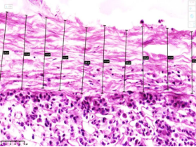
Figure 1. The average thickness of the pituitary gland capsule is presented. This micro-image was scanned using a NanoZoomer digital slide scanner. Digital values were extracted via NDP. view 2 software. H&E staining. Magnification: 10×10.
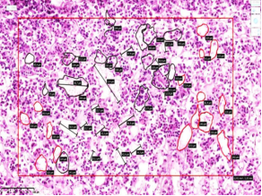
Figure 2. The area of acidophilic cell clusters within the pituitary gland structure is presented. This micro-image was scanned using a NanoZoomer digital slide scanner. Digital values were extracted via NDP.view 2 software. H&E staining. Magnification: 10×10.
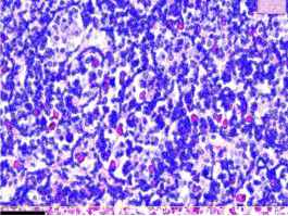
Figure 3. The cellular composition, total occupied area, and nucleocytoplasmic ratio within the pituitary gland parenchyma are presented. This micro-image was scanned using a NanoZoomer digital slide scanner. Digital values were extracted via QuPath-0.5.0 + NDP.view 2 software applications. H&E staining. Magnification: 10×10.

Information



 Share This Article
 Twitter
 Linked In
 Facebook
 Copy
Copy Download